Management of Thromboembolic Disorders in Vascular Surgery
Thromboembolic disorders, such as deep vein thrombosis (DVT) and pulmonary embolism (PE), pose significant risks to patients undergoing vascular surgery. Vascular surgeons and healthcare teams must employ comprehensive strategies for both prevention and management to minimize these potentially life-threatening complications. This article explores the current approaches and best practices in the management of thromboembolic disorders by vascular surgeon Silver Spring in vascular surgery.
Preoperative Risk Assessment
- Patient Evaluation: Comprehensive preoperative assessment is crucial. Patients should be evaluated for risk factors, including a history of DVT/PE, hypercoagulable conditions, malignancy, obesity, immobility, and advanced age
- Anticoagulant Medication Review: Review the patient’s medication history, including anticoagulant and antiplatelet agents. Determine the optimal management plan, which may include discontinuation or bridging therapy with low-molecular-weight heparin (LMWH) or direct oral anticoagulants (DOACs).
- Thrombophilia Screening: In select cases, screening for thrombophilic disorders may be warranted to identify underlying hypercoagulable conditions.
Prevention Strategies
- Pharmacological Prophylaxis: In patients at moderate to high risk of DVT/PE, prophylactic anticoagulation is often indicated. LMWH, unfractionated heparin (UFH), or DOACs may be used, balancing the risk of bleeding against thrombotic risk.
- Mechanical Prophylaxis: In patients at high risk of bleeding or contraindications to pharmacological prophylaxis, mechanical methods such as intermittent pneumatic compression (IPC) devices or graduated compression stockings (GCS) should be considered.
Center for Vascular Medicine – Silver Spring
10750 Columbia Pike Ste 605, Silver Spring, Maryland, 20901
(301) 396-7618
Intraoperative Measures
- Sequential Compression Devices (SCDs): Utilize SCDs during the surgical procedure, as they can help prevent venous stasis and lower extremity DVT.
- Anesthesia Considerations: Collaborate with anesthesiologists to maintain appropriate anesthetic depth, as deep anesthesia can increase the risk of thrombosis.
- Early Ambulation: Encourage early postoperative ambulation to reduce the risk of stasis and DVT formation.
Management of Thromboembolic Events
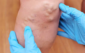
- Early Recognition: Train healthcare teams to recognize the signs and symptoms of DVT and PE, including leg swelling, pain, erythema, dyspnea, chest pain, and hemoptysis.
- Diagnostic Imaging: Employ diagnostic imaging, such as duplex ultrasound for DVT and computed tomography pulmonary angiography (CTPA) for PE, to confirm the diagnosis promptly.
- Anticoagulation: Initiate therapeutic anticoagulation with LMWH, UFH, or DOACs as soon as the diagnosis is confirmed. Tailor the choice of anticoagulant based on patient factors and the extent of thromboembolism.
- Thrombectomy and Thrombolysis: In select cases of massive PE or iliofemoral DVT, consider interventional procedures such as catheter-directed thrombolysis or thrombectomy.
- Vena Cava Filters: In patients with contraindications to anticoagulation or recurrent PE despite anticoagulation, consider placement of a vena cava filter to prevent further emboli.
- Long-term Anticoagulation: Develop a plan for long-term